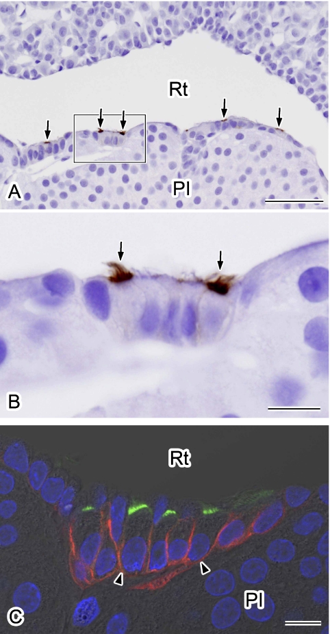
Fig. 4.
Immunolocalization of AQP5 in marginal layer cells. Paraffin sections were immunolabeled for AQP5 using an immunoperoxidase method (A, B). Nuclei were stained with hematoxylin. A: Labeling for AQP5 is evident in the apical surface of the marginal layer cells (arrows). B: A higher magnification view of the outlined area in A. AQP5 is localized at the apical membrane of some ciliated cells (arrows). C: Double-immunofluorescence staining for AQP5 (green) and AQP4 (red). Nuclei were stained with DAPI (blue). A single confocal image overlaid with differential interference-contrast image is shown. AQP5-positive cells are positive for AQP4 in their basolateral membranes (arrowheads). PI, pars intermedia; Rt, Rathke’s residual pouch. Bars=50 µm (A); 10 µm (B and C).

