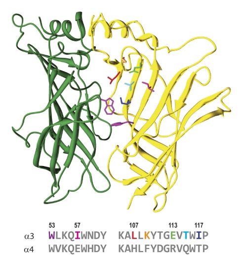
Fig. 4.
The noncanonical interfaces of α3β2 and α4β2 diverge. A homology model of the extracellular domain β2(+)/α3(−) interface is shown in ribbon format; β2 (left) is green and α3 (right) is gold. The tryptophan side chains (magenta; Trp149 of β2 and Trp53 of α3) conserved in the canonical/agonist binding site are shown in stick format. Below the structure is an alignment of the rat α3 and α4 sequences in the regions of the D and E loops. The side chains (stick format) chosen for specificity studies are color-coded in the α3 sequence; numbering is for the mature α3 protein. This depiction is derived from a3b2rr.pdb (Sallette et al., 2004).

