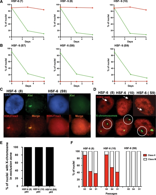
Figure 2.
Epigenetic status of the X chromosome with differentiation or serial passaging. (A) Six-day differentiation of HSF-6 (7), HSF-6 (8) and HSF-6 (10). OCT4+ and H3K27me3+ nuclei were counted at days 0, 3 and 6 of differentiation in each subline (n = 200 nuclei at each time point in each subline). (B) HSF-6 (S7), HSF-6 (S8), HSF-6 (S9) were differentiated in the same conditions as above (n = 200 nuclei for each data point). (C) XIST RNA FISH combined with immunostaining for H3K27me3. HSF-6 (8) (n = 80/80) nuclei exhibited co-localization of XIST with H3K27me3. HSF-6 (S9) (n = 120/120) nuclei did not exhibit any XIST signal. (D) X chromosome FISH combined with immunostaining for H3K4me3 (H3K4me3/X FISH). White and green arrows show nuclear exclusion zones. The white circles show the localization of one X chromosome in one exclusion zone. (E) Quantification of data in (D) (n = 10) for each subline. (F) Transition of class II to class III is unidirectional. Class II and class III nuclei were evaluated on duplicate cover slips collected at passages 65, 84 and 91 in HSF-6 (8), HSF-6 (10) and HSF-6 (S9). Class II nuclei were evaluated by scoring H3K27me3 foci (n = 200); XCI was identified using H3K4me3/X FISH (n = 10). Class III = n(H3K4me3/X FISH) – n(H3K27me3).

