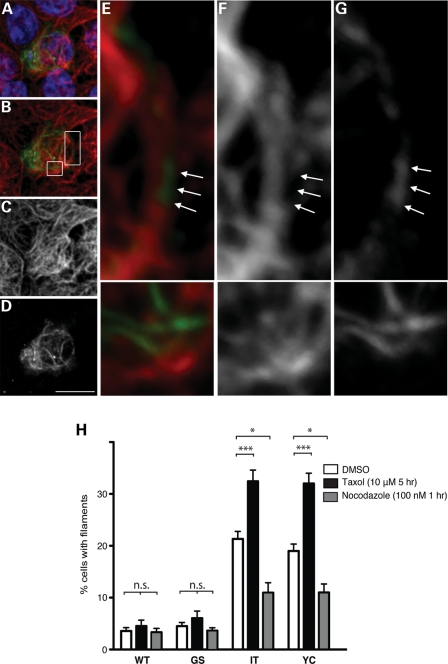
Figure 4.
LRRK2 filaments associate with microtubules and are modified by microtubule-altering drugs. (A–G) LRRK2 filaments localize to microtubules. HEK293T cells transfected with FLAG-LRRK2-I2020T were double labeled with anti-FLAG (D and G) and anti-alpha tubulin (C and F) antibodies. LRRK2 filaments appear as continuations of microtubules (A, B and E, merged images showing tubulin in red, LRRK2-FLAG in green and nuclei in blue; insets in B are shown in E, F and G). Arrows denote portions of microtubules positive for LRRK2 staining, but lacking tubulin staining. Scale bar is 10 μm. (H) Treatment with microtubule-altering drugs changed the filament proportion. CAD cells were transfected with WT or PD mutant forms (G2019S, I2020T or Y1699C) of GFP-LRRK2. Forty-eight hours post-transfection, cells were treated with the microtubule-stabilizing drug taxol (10 μm for 5 h), the microtubule-destabilizing drug nocodazole (100 nm for 1 h) or dimethyl sulfoxide (DMSO) control. Following drug treatment, cells were fixed, stained for GFP and the frequency of cells with filaments was quantified. Data are means ± SE of three independent experiments. (*P< 0.05, ***P< 0.001, n.s., non-significant; two-way analysis of variance (ANOVA) with post-hoc Tukey's test).

