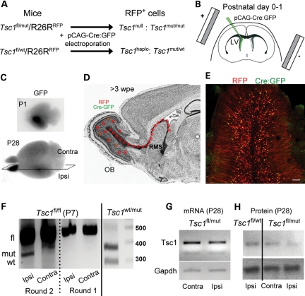
Figure 2.
Postnatal deletion of Tsc1 using neonatal electroporation and the Cre-Lox system. (A) Schematic describing the loss of one or two Tsc1 alleles in cells containing a plasmid encoding Cre:GFP under the CAG promoter that is expressed through neonatal electroporation in Tsc1fl/wt/R26R and Tsc1fl/mut/R26R, respectively. Cells containing the pCAG-Cre:GFP also express RFP. (B) The diagram illustrating the principle of neonatal electroporation of a plasmid into neural progenitor cells lining the lateral ventricle. (C) Images of P1- and P28 fixed brains containing RFP+ cells in the ipsilateral olfactory bulb. (D) The diagram illustrating the migratory path and the final location of newly born cells 3 weeks post-electroporation (wpe). RFP expression persists permanently due to its genomic integration (R26R mice), while GFP expression from either a pCAG-GFP or pCAG-Cre:GFP is diluted in newly born cells due to successive cell division. As a result, only cells born during the first 7–10 days express GFP. (E) The confocal image of RFP+ and GFP+ newly born cells in a coronal olfactory bulb section. RFP+/GFP+ cells outnumber RFP+/GFP− cells. Scale bar: 70 µm. (F) PCR gels from genomic DNA obtained from the ipsilateral (mRFP-containing cells) and contralateral olfactory bulb from P7 Tsc1fl/fl/R26R and Tsc1wt/mut/R26R mice electroporated at P1. (G) PCR gels of Tsc1 and Gapdh cDNA obtained from the ipsilateral and contralateral P28 Tsc1fl/mut/R26R olfactory bulb. All the gels were run on the same blots. (H) Western blots of TSC1 (hamartin) and GAPDH obtained from ipsilateral P28 Tsc1fl/wt/R26R and Tsc1fl/mut/R26R olfactory bulbs.

