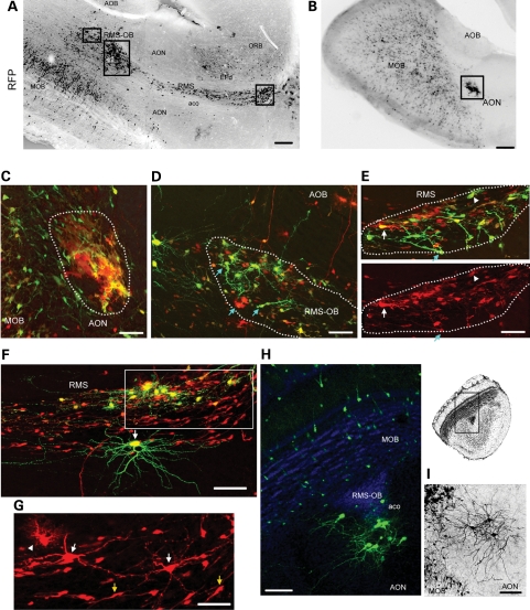
Figure 4.
Tsc1null cells form the migratory heterotopia in and out of the RMS. Low magnification photographs of RMS/RMS-OB (A) and AON (B) lesions containing RFP+ cells in sagittal sections of Tsc1fl/mut/R26R mice electroporated at P1 with pCAG-Cre:GFP and pCAG-GFP. The black rectangles illustrate the approximate locations of images shown in (C)–(E). (C) The confocal image of a lesion in the AON, the location of which is shown in (B). The lesion contains RFP+/GFP+ and RFP+/GFP− cells. (D–G) Confocal Z-stack images of lesions in the RMS-OB (D) and RMS (E and F), the locations of which are shown in (A). G is a zoom and smaller Z-stack of the region in the white rectangle in F. Blue arrows (D and E) point to cells with an immature morphology. White arrows (E, F and G) point to some neurons. The arrowhead (E and G) points to examples of cells resembling astrocytes. Yellow arrows (in G) point to neuroblasts. (H and I) Confocal Z-stack images of neuronal heterotopias in the aco of the olfactory bulb in a coronal section (GFP+ neurons in green and DAPI in blue, H) and the neuronal heterotopia in the AON (black, I). Scale bars: 700 µm (A and B), 40 µm (C), 35 µm (D), 50 µm (E and G) and 70 µm (F, H and I). aco, anterior commissure, olfactory limb; AOB, accessory olfactory bulb; AON, accessory olfactory nucleus; aco:EP, doral endopiriform cortex; ORB, orbital cortex.

