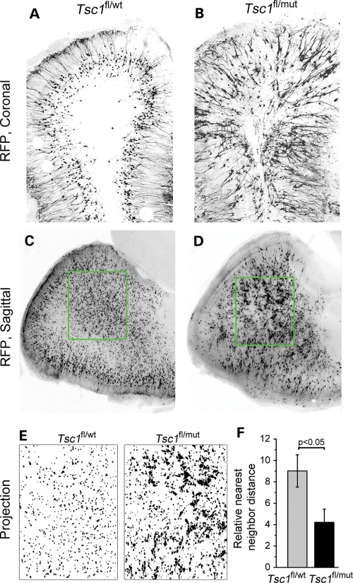
Figure 6.
Tsc1null neurons form micronodules throughout the olfactory bulb. Confocal images of RFP+ cells in olfactory bulb coronal (A and B) and sagittal (C and D) sections from Tsc1fl/wt/R26R (A and C) and Tsc1fl/mut/R26R mice (B and D). (E) Projections of the RFP signals in the green square in (C) and (D). (F) The bar graph of the relative nearest neighbor distance for RFP+ cells in the olfactory bulb of Tsc1fl/wt/R26R (gray) and Tsc1fl/mut/R26R (black) mice. Scale bars: 140 µm (A and B) and 200 µm (C and D).

