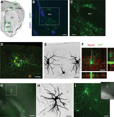
Figure 8.
Newborn Tsc1null neurons are rerouted to cortical and subcortical areas. (A) Coronal sections with green rectangles indicating the approximate locations of images shown in (B)–(I). (B) The confocal image of GFP+ neurons (green) in the ACB and around the aco counterstained with DAPI (blue). (C) The zoom of the image in the white square in (B). (D) The confocal image of GFP+ and RFP+ neurons and astrocytes (bushy cells) in cortical layer II. (E) GFP+ (black) neurons in cortical layer II. (F) The confocal image and projections of a GFP+ cortical layer II cell that immunostained positive for NeuN (red). (G) The confocal image of GFP+ neuron in the deep layer of the cortex. (H) Zoom of the neuron in the white square in (G). (I) The confocal image of GFP+ neuron in the deep layer of the cortex. Inset: GFP fluorescence overlaid with DIC to illustrate the location of the corpus callosum. CC, corpus callosum; CP, caudate putamen; ACB, accumbens nucleus; aco, anterior commissure. Scale bars: 140 µm (B) 70 µm (C), 80 µm (D and G), 40 µm (E, F and I) and 30 µm (H).

