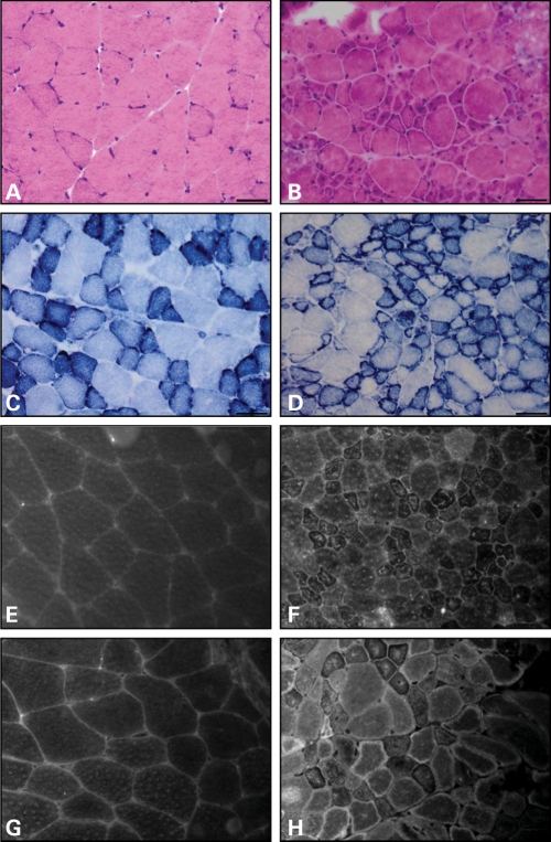
Figure 3.
Mtm1 p.R69C muscle develops structural changes of MTM. Hematoxylin and eosin-stained frozen cross section of quadriceps from 3-month-old wild-type (A) and Mtm1 p.R69C (B). Note the increased number of small myofibers, many of which show central nuclei in Mtm1 p.R69C animals. NADH-TR stain shows the expected delicate staining pattern in wild-type animals (C). In Mtm1 p.R69C muscle, (D) increased NADH-TR staining is noted at the periphery of small fibers. Quadriceps from a 3-month-old wild-type littermate (E, G) and an Mtm1 p.R69C mouse (F, H) stained with anti-DHPR antibodies to highlight T-tubules, and anti-RyR antibodies to mark sarcoplasmic reticulum. DHPR and RyR immunolocalization in wild-type animals show the expected delicate reticular staining pattern, while numerous fibers in Mtm1 p.R69C animals display alterations in the staining pattern including increase in central and peripheral staining, especially of small fibers. Bars = 50 μm.

