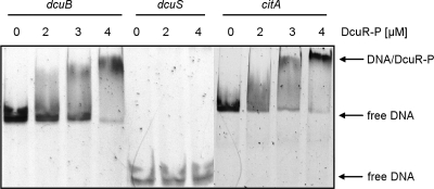
Fig 1.
Gel retardation of dcuB, dcuS, and citA promoter DNA by phosphorylated DcuR (DcuR-P). The promoter fragments of citA (652 bp), dcuB (603 bp), and dcuS (220 bp) were incubated in the presence of a 300-fold excess of competitor DNA with increasing concentrations of His6-DcuR-P as indicated. The protein-DNA mixture was subjected to nondenaturing DNA PAGE. The locations of the free promoter DNA and of the retarded DNA/DcuR-P complexes are indicated by arrows.

