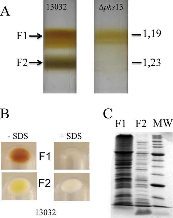
Fig 1.

Fractionation of C. glutamicum cell envelope by isopycnic sucrose gradient centrifugation. After cell lysis, membrane-containing fractions were separated according to density on a sucrose step gradient. (A) Centrifugation tubes for the wild-type 13032 strain and for its isogenic Δpks13 mutant. Buoyant densities (d) are indicated in g cm−3. (B) Fractions that correspond to F1 and F2, isolated from the 13032 strain, were pooled, treated with 4% SDS, and heated at 100°C for 30 min or left untreated. Insoluble material was collected by ultracentrifugation, and the corresponding pellets are shown. (C) SDS-PAGE protein pattern of F1 and F2 isolated from strain 13032 after Coomassie blue staining. The same amounts of F1 and F2, corresponding to approximately 5 × 10⁸ cells, were loaded on the gel. MW, molecular mass markers. The molecular masses of the bands with the highest intensities are 66 and 27 kDa, respectively.
