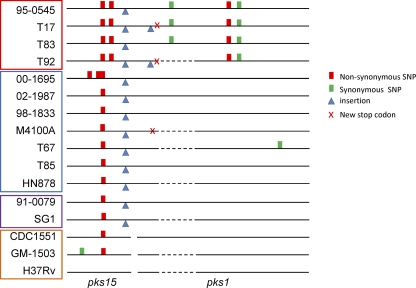
Fig 4.
Polymorphisms in pks15/1 loci of M. tuberculosis clinical strains and H37Rv. Sequences are compared to the published H37Rv sequence. Separate pks15 and pks1 loci are represented by a break in the black line. Dashed lines show regions that were unable to be sequenced in this study. Strains are highlighted to represent different M. tuberculosis clades: red, clade 1; blue, clade 2; purple, clade 3; orange, clade 4.

