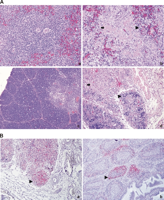
Fig 4.
Histological changes and distribution of Newcastle disease virus nucleoprotein in NDV-Peru/08-infected tissues. (A) Histological changes (panels b and d) consisted of lymphoid depletion and necrosis (arrows), accumulation of macrophages (arrowheads), and scattered heterophils. Panel a, spleen, PBS control, 40×; panel b, spleen, NDV-Peru/08, 40×; panel c, thymus, PBS control, 20×; panel d, thymus, NDV-Peru/08, hematoxylin and eosin staining. (B) Immunohistochemical staining for Newcastle disease virus nucleoprotein. Shown is positive staining in the thymus (panel a, 20×) and bursa of Fabricius (panel b, 20×). Positive cells consisted mainly of lymphocytes and macrophages (arrowheads).

