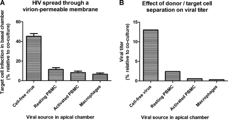
Fig 5.
Relative importance of cell-to-cell spread in a coculture of R5M target cells with HIV-infected effector cells. (A) Cell-free HIV or HIV-infected effector cells (x axis) were physically separated from the R5M target cells using a virion-permeable membrane with 3-μm pores in a 24-well Transwell system with the cell-free virus or effector cells placed in the apical compartment and the target cells in the basal compartment. In parallel, cocultures of cell-free virus or effector cells with R5M target cells were set up in the basal compartment. After 5 days of incubation, R5M infection was assessed using firefly luciferase. Values for target cell infection by cell-free HIV or HIV-infected effector cells when separated from the target cells are expressed as percentages of those obtained in the respective cocultures (y axis). Mean percentages ± standard errors of the mean (SEM) of three independent experiments are depicted. (B) Additionally, the TCID50 of the infected effector cells was determined as described in the legend to Fig. 2 using a 96-well Transwell system with the effector cells placed in the apical compartment and the target cells in the basal compartment. Viral titers were calculated using the method of Reed and Muench and expressed as percentages of the titer obtained in cocultures of effector and target cells. One experiment is represented.

