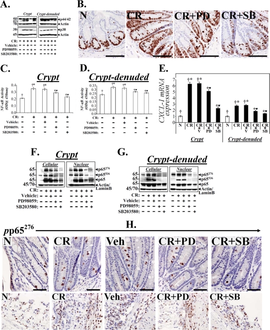
Fig 6.
Effect of p44/42-ERK1/2 and p38 inhibition on cell proliferation and NF-κB activity in vivo. Uninfected or C. rodentium-infected-infected C3H mice were injected once a day for 10 days with either vehicle or inhibitors specific for ERK1/2 (PD) or p38 (SB). At 2 h after the last injection, colonic crypts and CLP were isolated. (A) Representative Western blots showing total ERK1/2 and p38 in the two compartments. (B) Representative photomicrographs of paraffin-embedded sections stained with antibody to Ki-67: N, uninfected healthy mice; CR, C. rodentium-infected mice; CR+PD or CR+SB, C. rodentium-infected mice treated with specific ERK1/2 or p38 inhibitor. Scale bar = 50 μm (n = 3 independent experiments). (C and D) NF-κB activities measured via DNA binding assay in the crypts and CLP. Each bar represents mean ± standard deviation. †*, P < 0.05 versus control (†); *♥, P < 0.05 versus CR (†*) (n = 3). (E) Real-time RT-PCR. CXCL-1 expression in the crypts and CLP were measured via real-time RT-PCR. †*, P < 0.05 versus control (†); *♥, P < 0.05 versus C. rodentium-infected, vehicle-treated mice (CR+V; †*) (n = 3 independent experiments). (F and G) Crypts and CLP cellular and nuclear extracts prepared from the distal colons of the above group of animals were analyzed by Western blotting with antibodies specific for total p65 or the p65 subunit phosphorylated at Ser 276 (p65276) or Ser 536 (p65536). Actin or lamin B was the loading control, respectively (n = 3 independent experiments). (H) Representative photomicrographs of paraffin-embedded sections stained with antibody specific for phosphorylated p65276 (pp65276): N, uninfected healthy; CR, C. rodentium-infected; Veh, C. rodentium-infected and vehicle-treated; CR+PD or CR+SB, C. rodentium-infected and treated with specific ERK1/2 or p38 inhibitor, respectively. Scale bar = 30 μm (n = 3 independent experiments).

