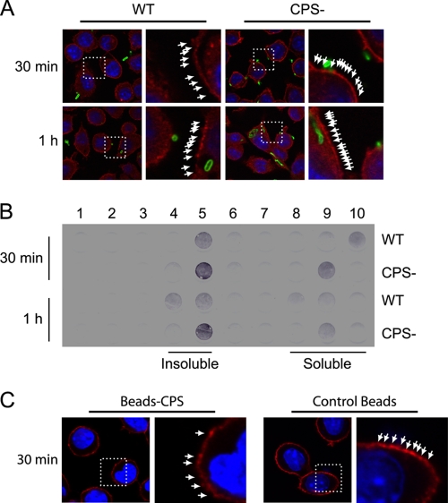
Fig 4.
CPS disrupts lipid microdomains during phagocytosis of S. suis by macrophages. J774 macrophages were infected with S. suis WT or its nonencapsulated mutant strain (CPS−) for 30 min and 1 h. (A) Confocal analysis of lipid microdomain distribution. Cells were stained with fluorescent cholera toxin (red), which binds to GM1 ganglioside in lipid microdomains, and with rabbit anti-S. suis antibodies (green) to stain bacteria. The pattern of staining of GM1 (arrows) is more diffuse at 30 min and 1 h when cells are infected with WT S. suis than when they are infected with the CPS− mutant. (B) Dot blot analysis of lipid microdomain distribution. Cells were infected as described above and lysed, and membranes were fractionated by ultracentrifugation on an OptiPrep gradient (1, top fraction; 10, bottom fraction). GM1 ganglioside was revealed by incubating with cholera toxin-HRP. Insoluble membrane fractions corresponding to lipid microdomains are shown in fractions 4 and 5. Soluble membrane fractions are shown in fractions 8, 9, and 10. Macrophages showed a higher GM1 label intensity in lipid microdomains when infected by the CPS− mutant than in cells infected by WT S. suis. (C) Confocal analysis of lipid microdomain distribution after macrophages were incubated with either unlinked control beads or CPS-linked beads (Beads-CPS) for 30 min. Cells were stained with fluorescent cholera toxin (red). The pattern of staining of GM1 (arrows) is more diffuse when cells are treated with CPS-linked beads than when they are treated with control beads.

