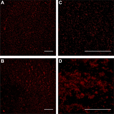
Fig 5.
Fluorescent micrographs of OG1RF (A and C) and Δeep strain (B and D) biofilms at 6 h postinoculation. Biofilms were grown on Aclar fluoropolymer coupons in tryptic soy broth without added dextrose, washed, and stained with wheat germ agglutinin conjugated to Alexa Fluor 594 as described in Materials and Methods. Representative images acquired at magnifications of ×200 (A and B) and ×600 (C and D) are shown. Scale bars, 20 μm.

