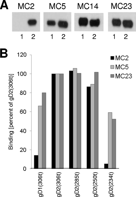
Fig 2.
Mapping of antibody epitopes on gD for the MC MAbs. (A) Western blot analysis of MC2, MC5, MC14, and MC23. Purified truncated gD1(306), indicated as 1, or gD2(306), indicated as 2, were subjected to SDS-PAGE under nondenaturing conditions. Western blots were probed with the indicated MAbs. (B) ELISA. Truncated gD1(306t), as well as gD2 truncated at amino acid 306, 285, 250, or 234, was reacted with the same IgG concentration of MC2, MC5, or MC23 for 1 h at RT. Bound antibody was detected by an anti-mouse secondary antibody and ABTS [2,2′-azino-bis(3-ethylbenzothiazoline-6-sulphonic acid)] substrate. Absorbance values were normalized by setting the values obtained for each MAb binding to gD2(306t) as 100%.

