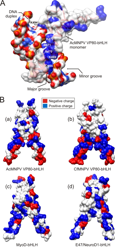
Fig 6.
Surface models of VP80-bHLH domains. (A) Surface model of AcMNPV VP80-bHLH (monomer) in complex with a double-stranded DNA molecule. The distribution of positively (blue) charged residues within the basic region (K466, K469, R470, R471, K472, and K494) of bHLH perfectly matches the major groove of DNA duplex flanked by negatively (red) charged sugar-phosphate backbone. (B) Gallery of bHLH dimer structures for AcMNPV-VP80 (a), CfMNPV-VP80 (b), human MyoD (c), and E47/NeuroD1 (d). Surface models show the distribution of positive (blue) and negative (red) charged residues. Typical for baculovirus VP80-bHLH domains is the presence of negatively (red) charged residues at the proximal domain ends.

