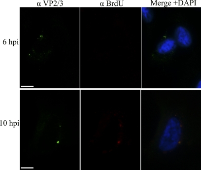
Fig 8.
Exposure of VP2/3 precedes exposure of the genomic DNA. Representative 6- and 10-h samples from the experiment summarized in Fig. 7 are shown. Samples were stained for BrdU (Cy3) and VP2/3 (FITC), and the nucleus was stained with DAPI (blue). Scale bar, 10 μm.

