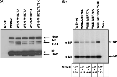
Fig 2.
Viral proteins in virus-infected cells and virions. (A) Expression of viral proteins in virus-infected cells. MDCK cells were infected with the indicated viruses at an MOI of 5 and were grown in the presence of TPCK-trypsin. Cytoplasmic extracts were prepared at 8 h postinfection and analyzed by use of Western blotting with a polyclonal antibody against WSN virus. Arrows to the right indicate the positions of the viral proteins. (B) Viral proteins in virions. Viruses grown in MDCK cells in the presence of TPCK-trypsin were concentrated by ultracentrifugation through a sucrose cushion and resuspended in RIPA buffer. The protein samples were then analyzed by means of Western blotting with either anti-NP or anti-M1 monoclonal antibody. The intensity of bands for NP and M1 was quantitated by using ImageJ software (NIH). The NP/M1 quantitative ratio was calculated (the value for the wild-type virus was taken as 1), and the averages from three independent experiments (with standard deviations) are shown at the bottom of the gel image. Arrows to the right indicate the position of the viral proteins.

