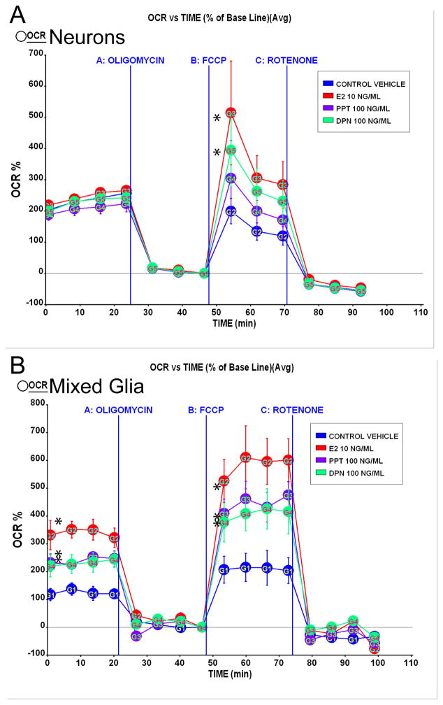
Figure 7. SERMs increased mitochondrial respiration in vitro.
A. Primary rat embryonic (E18) neurones from hippocampus were cultured for 10 days in Neurobasal Media with B27 supplement and then treated with E2 (10 ng/ml), PPT (100 ng/ml), or DPN (100 ng/ml) or vehicle for 24 h prior to the experiment. Oxygen consumption rates (OCR) were determined using Seahorse XF-24 Metabolic Flux Analyzer. Vertical lines indicate time of addition of mitochondrial inhibitors A: oligomycin (1 μM), B: FCCP (1 μM) or C: rotenone (1 μM). OCR in primary neurones treated with E2 (10 ng/ml) and DPN (100 ng/ml) exhibited a greater magnitude of maximal mitochondrial respiratory capacity compared to vehicle (*=P<0.05 vs. vehicle; n=4). Primary rat embryonic (E18) mixed glia from hippocampus were cultured for 14 days in DMEM supplemented with 10% serum. B. Glial cells derived from primary rat embryonic (E18) hippocampus were cultured for 10 days before re-plating. Mixed glial cells were serum-starved 4 h then treated with E2 (10 ng/ml), PPT (100 ng/ml), DPN (100 ng/ml) or vehicle for 24 h prior to the experiment. Vertical lines indicate time of addition of mitochondrial inhibitors A: oligomycin (1 μM), B: FCCP (1 μM) or C: rotenone (1 μM). OCR in primary glia treated with E2 (10 ng/ml), PPT (100 ng/ml), or DPN (100 ng/ml) displayed greater basal respiration and increased maximal mitochondrial respiratory capacity compared to vehicle (*=P<0.05 vs. vehicle; n=4).

