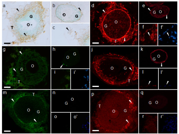
Figure 6.

Immunohistochemical localization of the theca cell markers in cultured follicles. Immunohistochemistry of (a-c) fibronectin, (d-f) laminin, (g-i) tenascin, (j-l) collagen type IV, (m-o) THY1, and (p-r) CYP17A-1. (f, f'), (i, i'), (l, l'), (o, o'), and (r, r'), are same view respectively. (f', i', l', o', r') are DAPI staining. (d-r') visualized by immunofluorescence and (a-c) visualized by ABC staining. (a, d, g, j, m, p) were cultured follicles for 5 days, (b, e, h, k, n, q) were cultured follicles for 0 day (isolated follicle), and (c, f, f', i, i', l, l', o, o', r, r') were interstitial cells distanced from follicles. The arrowheads indicate each signal. O, oocyte; G, granulosa cells; T, theca cells. Scale bars = 25 μm.
