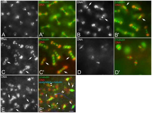
Figure 2. Improper chromosome segregation during the intravitelline mitoses.
Black and white panels represent DNA staining alone; colour panels represent microtubules (green), DNA (red), and the sperm tail (blue). (A,A′) Detail of a normal embryo during early telophase of the seventh nuclear division cycle: sister chromatids have migrated at the opposite poles of the spindles to form daughter nuclei. Detail of embryos during early telophase (B,B′) or late anaphase (C,C′) of the seventh mitosis showing abnormal spindles: sister chromatids either do not separate and stay at the spindle equator (arrows in B) and the spindles are barrel-shaped (arrows in B′) or begin to separate, but do not migrate properly lagging in the spindle midzone or forming chromatin bridges (arrows in C), spindles do not show apparent defects at this stage (arrows in C′). (D,D′) Unfertilized eggs fixed 1 hour AED: note the anastral barrel spindles. (E,E′). Abnormal fertilized embryos fixed 1 hour AED: abnormal mitotic figures range from barrel-shaped biastral spindles (arrows in E′) to aberrant late telophase spindles in which the chromatin form dense bridges (larger arrows in E) and the centrosomes maintain their position closest to daughter nuclei (arrowheads in E′); late telophase spindles may be still recognized by the midbody remnants (small arrows in E′); double arrows in E′ point to the sperm tail. Bacteria associate with the poles of biastral spindles, but not with the poles of the anastral spindles. Scale bar is 4 µm.

