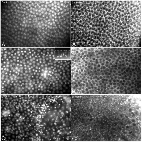
Figure 4. Nuclear condensation defects are amplified at cellularization.
Embryos are stained for DNA (left panels) and microtubules (right panels). Cellular blastoderm is usually formed by evenly spaced nuclei at the same condensation stage and of the same size and dimensions (A) surrounded by honeycomb microtubular baskets (A′). Abnormal cellular blastoderms are characterized by small (arrow in B) or larger (arrows in C) clusters of picnotic nuclei scattered among normal looking blastoderm nuclei; the honeycomb microtubular baskets are often altered in these embryos (arrowheads in B′, C′). Scale bar is 15 µm.

