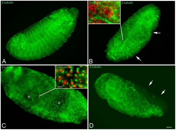
Figure 5. Embryos at later stages of development have highly defective areas.
Embryos were fixed after 15–20 hours AED and stained for microtubules (green) and DNA (red). (A) Normal developing embryos in which the shortening of germ band is completed: segments are well evident. (B,C) Defective embryos after the retraction of the germ band: (B) the integrity of some segments is affected (arrows) and (C) large areas of the embryos are incompletely formed and still showed mitotic divisions (asterisks). (D) Detail of an embryo during germ band shortening: note the lack of differentiation of the anterior region of the body (arrows). Insets are details of the surface areas. Scale bar is 25 µm in the main panels and 7 µm in insets.

