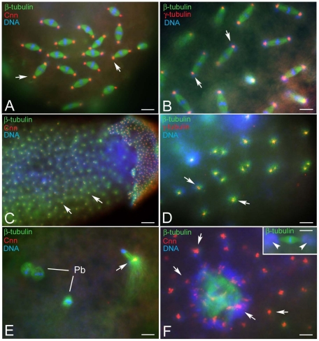
Figure 8. The focus of the cytoplasmic asters contains centrosomal components.
Fertilized (A,B) and unfertilized (C–F) eggs obtained by KOS10 females are incubated with antibodies against β-tubulin (green); Cnn and γ-tubulin (red) and counterstained with Hoechst 33258 (blue). Cnn and γ-tubulin are evident at the spindle poles of normal developing embryos and at the focus of the cytoplasmic asters (arrows, A–D). (E) Detail of an unfertilized oocyte at the end of meiosis with a cluster of centrosomal material within the remnant of the central aster (arrow); Pb, polar bodies. (F) Cnn aggregates are found within the cytoplasm (arrows) and around the meiotic-like spindles (F, arrows), but not at their poles (inset F, arrowheads), despite the presence of microtubule asters and associated bacteria. Bar is 25 µm in C and 4 µm in all other panels.

