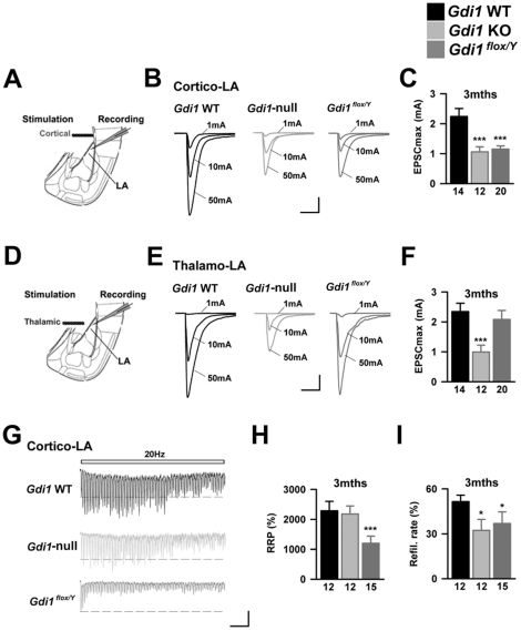
Figure 8. Delayed Gdi1 deletion leads to exaggerated synaptic phenotypes.
(A) Scheme of the slice preparation (B) Evoked EPSCs obtained in Gdi1 WT, Gdi1-null and Gdi1flox/Y for different stimulation intensities were recorded. Scale bars: 500 pA and 15 msec. (C) Bar graph presenting the mean EPSC amplitude at maximal stimulation intensity (50 mA*msec, defined as EPSCmax) in the various genotypes. Number of recorded cells is indicated. ***p<0.001. (D–F) Same presentation as in A–C for the examination of Thalamo-LA synapses. (G) Representative postsynaptic responses to high frequency trains of stimulation in 3 months old Gdi1 WT, Gdi1-null and Gdi1flox/Y animals. Scale bars: 50 pA and 500 msec. (H) Calculated RRP size was decreased in Gdi1flox/Y whereas normal in Gdi1-null mice. ***p<0.001 (I) The refilling rate was lowered in both null and conditional Gdi1 mutant mice. *p<0.05.

