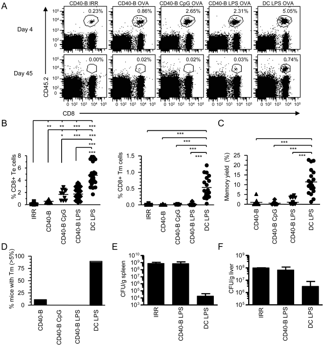
Figure 2. Immunization with CD40-B cells induces an in vivo CD8+ T cell response.
A. CD40-B cell vaccination generates CD8+ Te cells but not Tm cells. 106 female OT-1 T cells (CD8+CD45.2+) were adoptively transferred into congenic B6SJL female mice (CD45.1+) followed by immunization two days later with 2×10⁶ CD40-B cells, matured or not with LPS (1 µg/mL) or CpG (2 mM) and loaded with 4 µg/mL OVA or with an irrelevant peptide (IRR). As a reference recipients were immunized with 2×10⁶ DCs matured with LPS and loaded with OVA peptide. OVA-specific T cells (CD8+CD45.2+) were analyzed in the same mouse by surgical removal of superficial lymph nodes at d4 (effector) and d30–45 (memory) post-immunization. Te and Tm cells were identified as CD8+CD45.2+ by flow cytometry. The percentage of Te and Tm cells generated are indicated on each dot plot. B. Percentage of CD8+ Te (left panel) and Tm (right panel) cells recovered at d4 (Te) and d>30 (Tm) in one lymph node is shown. C. Yield of CD8+ Tm cell generation. The yield of Tm cell formation was calculated as the percentage of Te cells that develop into Tm cells. D. The percentage of mice that generates more then 5% of CD8+ Tm cells is shown for the different immunization conditions. E and F. Lm challenges. 30 d post immunization, mice were challenged with a lethal dose of Lm-OVA (105 CFU). 3 d post challenged, CFU were determined in the spleen (E) and liver (F) for each mouse. A–D are from at least four independent experiments with at least two mice per group while E and F are from one independent experiment with three mice per group. * p<0.05, ** p<0.01 and *** p<0.001.

