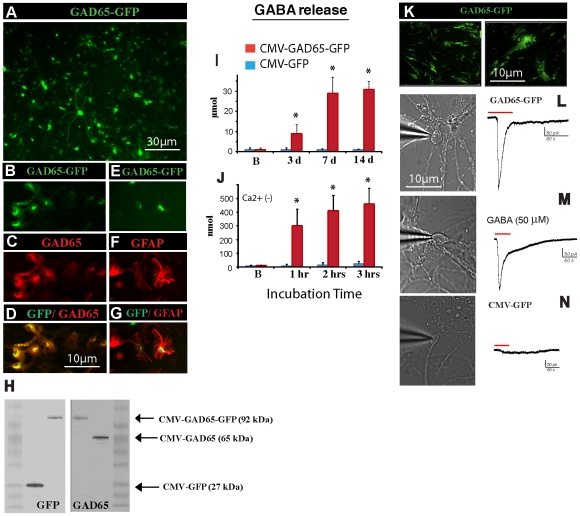
Figure 2. Infection of rat primary spinal cord culture with HIV1-CMV-GAD65 or HIV1-CMV-GAD65-GFP lentivirus leads to a preferential astrocyte GAD65 expression and release of biologically active GABA.
(A) Rat spinal cord primary culture infected with HIV1-CMV-GAD65-GFP lentivirus and stained with anti-GFP antibody at 4 days after lentivirus infection. (B–D) Co-staining of HIV1-CMV-GAD65-GFP-infected cells with GAD65 antibody showed preferential GAD65 expression in GFP-IR cells. (E–G) Colocalization of GFP-IR with GFAP-IR in HIV1-CMV-GAD65-GFP-infected astrocytes at 14 days after infection. (H) Western blotting for GFP or GAD65 in cell lysates taken from rat primary spinal cord culture infected with HIV1-CMV-GFP (control), HIV1-CMV-GAD65-GFP, and HIV1-CMV-GAD65 lentivirus. (I) Extracellular GABA release measured in cell culture media taken from rat primary spinal cord culture 3–14 days after HIV1-CMV-GFP (control) or HIV1-CMV-GAD65-GFP lentivirus injection. (J) Progressive increase in extracellular GABA release measured in Ca2+-free media 1–3 hrs after cell culture wash in HIV1-CMV-GAD65-GFP but not in HIV1-CMV-GFP (control) lentivirus-infected cells (* P<0.01; paired t test). (K) Human fetal spinal cord astrocytes infected with HIV1-CMV-GAD65-GFP lentivirus and stained with anti-GFP antibody at 7 days after lentivirus infection. (L) Changes in whole-cell inward current in cultured human NT neurons after bath application of human astrocyte-HIV1-CMV-GAD65-GFP-conditioned media, (M) 50 µM GABA or (N) human astrocyte-HIV1-CMV-GFP-conditioned media (control); (neurons clamped at holding potential (-) 60 mV).

