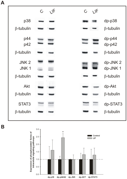
Figure 5. Analysis of intracellular signaling pathways that mediates LIF actions on lung growth.
(A) Western blot analysis of p38, p44/42, JNK1/2, Akt and STAT3, and to diphosphorylated forms of p38 (dp-p38), p44/42 (dp-p44/42), SAPK/JNK (dp-JNK1/2), Akt (dp-Akt) and STAT3 (dp-STAT3) in control lung explants (C) and treated with LIF at 40 ng/mL (LIF). Control loading was performed using β-tubulin (55 kDa). p38 corresponds to 38 kDa. p44/42 correspond to 44 and 42 kDa, respectively. JNK1 and 2 correspond to 46 and 54 kDa, respectively. Akt corresponds to 60 kDa. STAT3 corresponds to two bands, 79 and 86 kDa. (B) Semi-quantitative analysis of expression of phosphorylated forms of these intracellular signaling pathways. Results are presented as arbitrary units normalized for β-tubulin. p<0.05: * vs. control.

