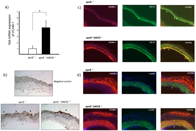
Figure 2. eNOS deletion increases VCAM-1 expression.
a) Real time PCR analysis showed four fold increased expression of VCAM-1 mRNA in apoE−/−/eNOS−/− (n = 9) carotids, compared to apoE−/− (n = 20, *p<0.01). b) Immunohistochemistry confirmed increased endothelial VCAM-1 expression in carotid arteries of apoE−/−/eNOS−/−, compared to apoE−/−. Arrows indicate positive DAB staining (internal carotid artery, location of IVM). c) Double immunofluorescence staining of VCAM-1 protein in atherosclerotic lesions. Sections of the aortic arch of apoE−/− and apoE−/−/eNOS−/− animals were incubated with anti-VCAM-1 antibody (red) and anti-CD31 antibody (endothelial cells, green). Arrows indicate localization of VCAM-1 in endothelial cells in the overlay (yellow). Increased endothelial expression of VCAM-1 was observed in apoE−/−/eNOS−/− compared to apoE−/−. d) Increased medial smooth muscle cell expression of VCAM-1 was observed in advanced plaques in the aortic arch in apoE−/−/eNOS−/− compared to apoE−/−, as shown in yellow (arrows) by the double immunofluorescence staining of VCAM-1 (red) and smooth muscle cells (green).

