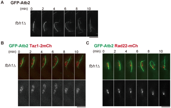
Figure 4. The fbh1 deletion mutant shows similar phenotypes to the skp1-a7 mutant.
(A) The bent spindle was frequently observed in fbh1Δ cells at MI. Time-lapse imaging for GFP-Atb2 is shown. (B) The fbh1Δ mutant exhibited non-disjunction of chromosome arms visualized by Taz1-2mCherry (red) with GFP-Atb2 (green). (C) The fbh1Δ mutant showed persistent Rad22-mCherry foci until MI. Bars, 5 µm.

