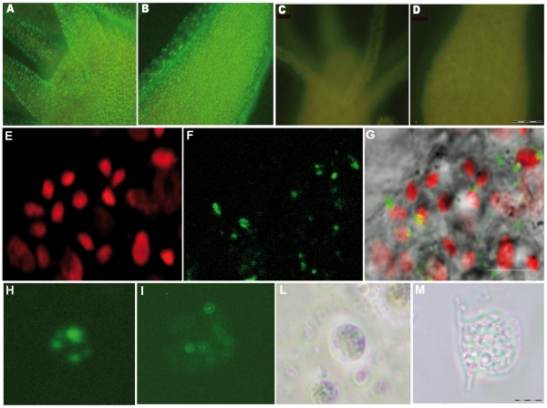
Figure 1. In vivo RNAi mediated by siRNA.
Fluorescence imaging of Hydra vulgaris exposed to myc-siRNA at different pHs. Living polyps were challenged with 70 nM Alexa488-end labelled myc-siRNA, in Hydra medium either at pH 4 (A, B) or at pH 7 (C, D). After 24 hr of continuous incubation with siRNA a strong punctuated fluorescence labels uniformly the whole animal, from the head (A) along the body (B) of animals treated at acidic pH, indicating the uptake of siRNA. In (C) and (D) are shown the body regions corresponding to A and B, respectively, of animals treated with the same siRNA at neutral pH. The absence of fluorescence signals indicates that the acidic pH enhances siRNA delivery in Hydra. Scale bar 200 µm. Confocal laser scanning imaging (E–G) of living Hydra treated 24 hr with Alexa488-labelled myc-siRNA revealed localization of oligonucleotides into interstitial cells. In (E), nuclear staining of interstitial cell nest with TOPO3; in (F) siRNA green fluorescence appears as faint staining or more evident granules; in (G), the overlay of bright field and fluorescence images revealed that siRNAs localize prevalently into the cytoplasm of interstitial cells. Scale bars: 10 µm. Fluorescence microscopy analysis of single cells suspensions prepared from treated dissociated animals shows intracellular localization of siRNA into interstitial (H) and epitheliomuscular (I) cells, as a punctuated pattern. The same cells were observed by phase contrast microscopy, respectively in (L) and (M). Scale bar 10 µm.

