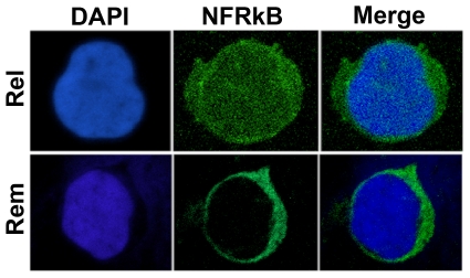
Figure 3. Confocal microscopy analysis of NFRKB staining in MCNS PBMC.
Immunofluorescence of PBMC from relapse and remission with anti-NFRKB antibody. Note that NFRKB is expressed in nuclei and cytoplasm compartments during the relapse but it is restricted to cytoplasm in remission. These data are representative of three independent experiments.

