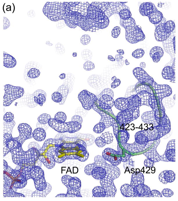
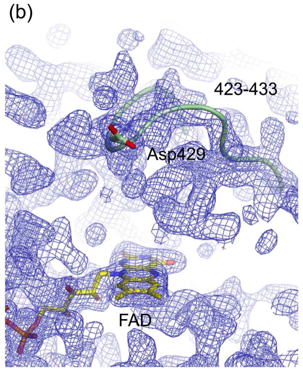
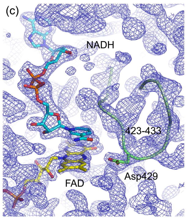
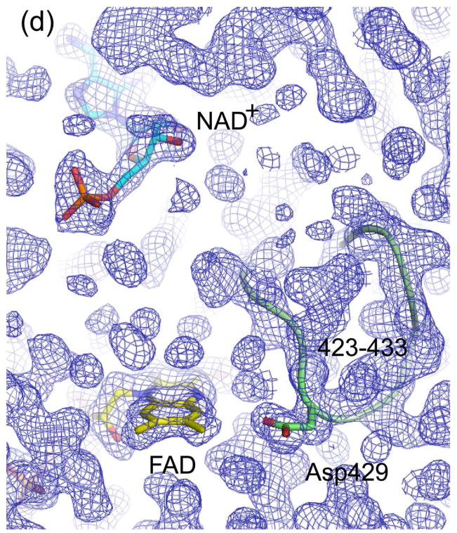
Figure 5.
Electron density maps of the FAD and NAD(H) binding sites in (a) ligand-free XDH, (b) ligand-free XO, (c) the XDH-NADH complex, and (d) the XDH-NAD+ complex. For clarity, only the FAD, NAD(H), and Asp429 are shown in atom representation. The backbone trace of the 423-433 loop is indicated by a green thread. Although the loop seems to swing away from the flavin ring in XO it does interfere with dinucleotide access in its new location.


