
Figure 7.
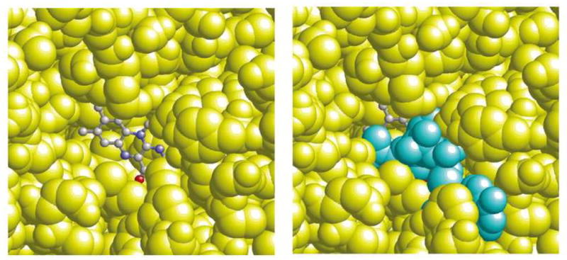
Representation of the FAD site of XDH. In the left hand figure, the NADH molecule has been removed to indicate the water-accessible area of the flavin ring. In the right hand figure, the same view is given but with the NADH molecule included to show how well the NADH molecule covers the flavin ring. The FAD atoms are depicted as ball-and-stick while the NADH molecule (cyan) and the protein atoms (yellow) are depicted as space-filling models.