
Figure 2. Skipping of exon 18 resulting from the splice donor IVS18+3insG mutation in case R09-068A.
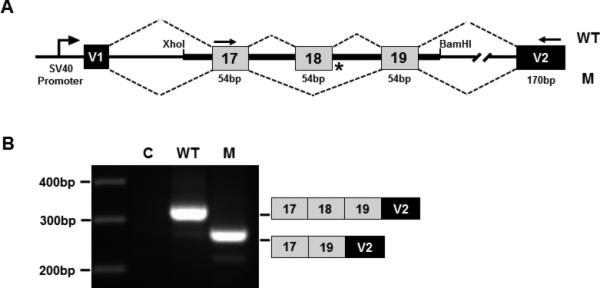
(A) Splicing construct showing the COL11A2 genomic region and vector/collagen exon organization. V1 and V2 are the vector exons. Normal and mutant COL11A2 genomic DNA fragments containing exons 17-19 were cloned into the XhoI and BamHI restriction endonuclease cleavage sites of the vector. (B) Gel electrophoresis image of fragments generated by RT-PCR from RNA derived from transfected cells using vector only (C), normal (WT) and mutant (M) constructs. Diagrammatic representations of the PCR product exon structures, based on sequence analysis, are shown on the right. PCR primers used to generate the fragments are indicated by arrows in (A).