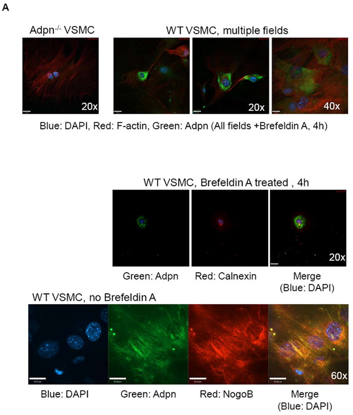
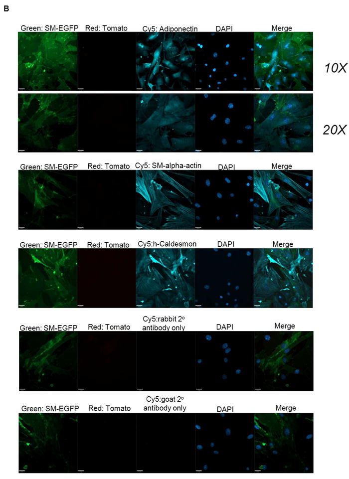
Figure 2. Adiponectin is synthesized in VSMC.


(A) Upper panel: VSMC from WT and adiponectin knock-out mice were treated with 100 ng/mL brefeldin A for 4 hours to arrest transport from ER to golgi to facilitiate visualization of ER proteins. Confocal microscopy was performed to show adiponectin (green), F-actin (red) and nuclei (DAPI, blue) staining, and multiple WT fields are shown. Middle panel: WT mouse VSMC treated with 100 ng/mL brefeldin A for 4 hours were stained for adiponectin (green), Calnexin (an ER marker, red), and DAPI (blue). Bottom panel: WT mouse VSMC (without brefeldin A treatment) were stained for adiponectin (green), NogoB (an ER protein, red), and DAPI (blue). The merged image (co-localization) is at right. (B) VSMC isolated from SM-GFP mice were stained for adiponectin, SM-alpha-actin, h-Caldesmon (blue, Cy5 secondary antibody), or fluorescent secondary antibodies only, and subjected to confocal microscopy analysis. Transgelin-lineage VSMC are green, non-SMC are red (tomato). Nuclei are stained with DAPI. No tomato red staining was detected.
