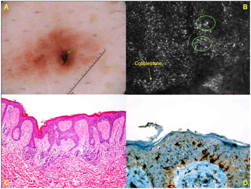
Case 2.

A. Dermoscopic image of a brown lesion (group A) on the abdomen of 61-year-old male shows an ill-demarcated lesion with a patchy network and an eccentric, peripheral black blotch. B. Basic RCM image (0.5mm × 0.5 mm) at the level of the spinous layer, from the dermoscopically-identified eccentric blotch, demonstrates a cobblestone pattern (→) in which few small, bright pleomorphic cells (❍) are seen in pagetoid pattern. C. Histopathologic analysis (H&E; original magnification ×50) shows a compound melanocytic nevus. There is mostly-lentiginous proliferation of melanocytes along the dermo-epidermal junction, but melanocytes in pagetoid pattern are not seen. D. Immunohistochemistry for CD1a reveals scattered intraepidermal Langerhans cells.
