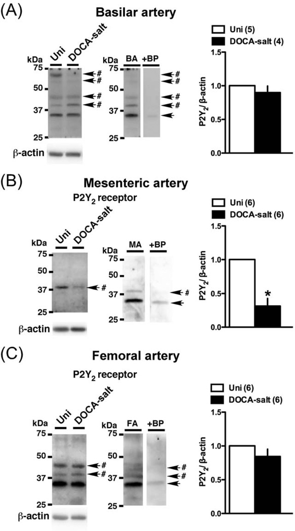
Fig. 7.
Western blots for P2Y2 receptor in basilar artery (A), mesenteric artery (B), and femoral artery (C) from DOCA-salt and Uni rats. Left: representative Western blots for P2Y2 and β-actin. #Band disappeared after the incubation of the primary antibody with the respective blocking peptide. BP, blocking peptide. Right: bands were quantified as described in Materials and methods. Ratios were calculated for the optical density of each receptor over that of β-actin. Number of determinations is shown within parentheses. *P < 0.05, DOCA-salt vs. Uni group.

