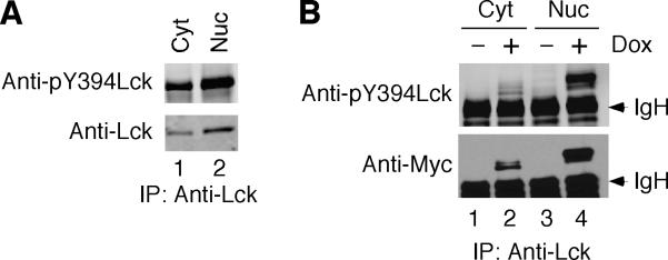
Fig. 3.
Phosphorylation of positive regulatory tyrosine of nuclear Lck. Cytosolic and nuclear fractions were isolated from LSTRA (A) and T-REx-293/Y505FLck (B) cells as described for Fig. 2. Normalized total proteins were immunoprecipitated with anti-Lck antibody, followed by immunoblotting with antibody specific for Tyr 394-phosphorylated Lck. The membrane was stripped and then reblotted for total Lck using anti-Lck (A) or anti-Myc tag (B) antibody. The arrowheads mark the positions of immunoglobulin heavy chain (IgH).

