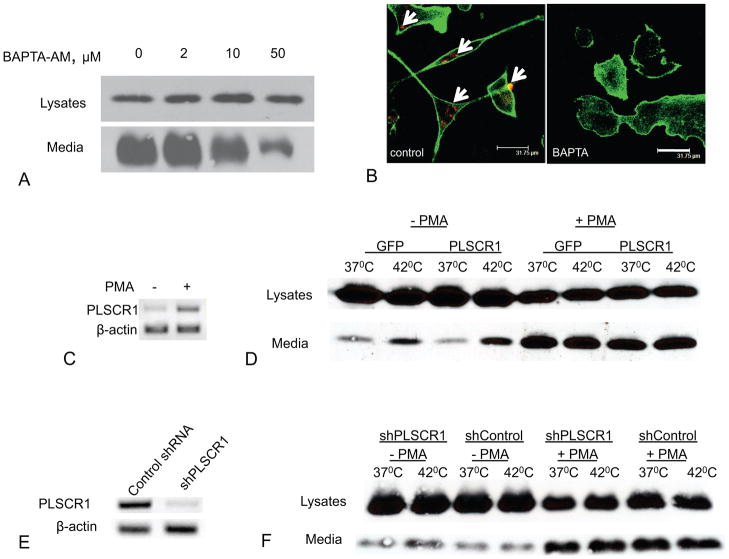
Figure 5. Intracellular calcium but not PLSCR1 is required for FGF1 export from U937 cells.
Each figure is representative of three independent experiments. (A). BAPTA-AM inhibits FGF1 export from U937 cells in a concentration-dependent manner. U937 cells stably transfected with FGF1 were treated with 150 nM PMA for 48 h, and then incubated for 110 min at 37°C in presence or absence of BAPTA-AM. FGF1 was isolated from conditioned media and detected using immunoblotting. 100% medium and 2% cell lysate were loaded per lane. (B). BAPTA-AM inhibits PS externalization in differentiated U937 cells. PMA-treated U937 cells were preincubated for 2 h with or without 50 μM BAPTA-AM and then incubated for 20 min with phycoerythrin-conjugated Annexin V (red), formalin fixed, permeabilized, stained with Alexa 488-phalloidin (green) and studied using confocal microscopy. PS externalization is marked with arrows. Bar – 31.75μm. (C). Undifferentiated U937 cells have low PLSCR1 expression, which is enhanced with PMA treatment. RNA was isolated from undifferentiated U937 cells and U937 cells treated for 48 h with 150 nM PMA. RT-PCR was performed using PLSCR1 and β-actin (control) primers. (D). Over-expression of PLSCR1 does not enhance FGF1 export from U937 cells. U937 cells stably transfected with PLSCR1:GFP or with GFP (control) were treated or not with 150 nM PMA for 48 h, and incubated for 110 min at 37°C or 42°C. FGF1 was isolated from conditioned media and detected by immunoblotting. 100% medium and 10% cell lysate were loaded per lane. (E). Specific shRNA decreases PLSCR1 expression in U937 cells. RNA was isolated from PMA-treated U937 cells stably transfected with PLSCR1 shRNA or control shRNA. RT PCR was performed using PLSCR1 and β-actin (control) primers. (F). shRNA knockdown of PLSCR1 does not affect FGF1 export from U937 cells. U937 cells stably transfected with FGF1 and cotransfected with PLSCR1 shRNA or control shRNA were treated or not with 150 nM PMA for 48 h, and then incubated for 110 min at 37°C or 42°C. FGF1 was isolated from conditioned media and detected using immunoblotting.

