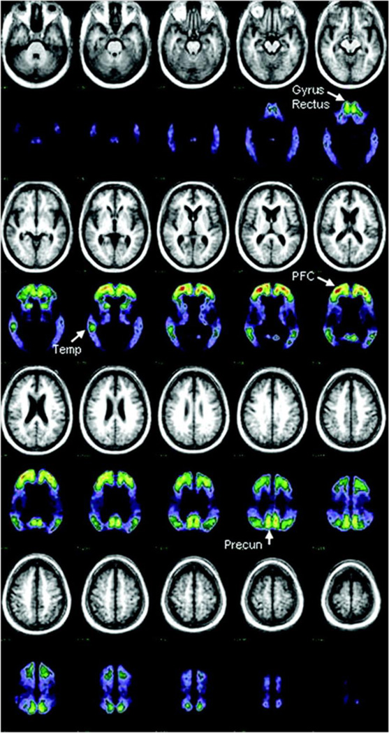
Figure 1.
Mean MRI and PET [11C]PIB distribution in a standard atlas coordinate system from 10 subjects with dementia of the Alzheimer type. PET data represent [11C]PIB activity in the late (30 to 60 minutes after injection) distribution and has been normalized to standardize display and increase contrast. Brain areas used to detect raised [11C] PIB uptake in nondemented subjects are indicated with arrows. PFC = prefrontal cortex; Temp = temporal cortex; Precun = precuneus region. Reproduced with permission from [84].

