
Figure 2. Keratin Organization is Regulated by Tension on Cell-Cell Contacts.
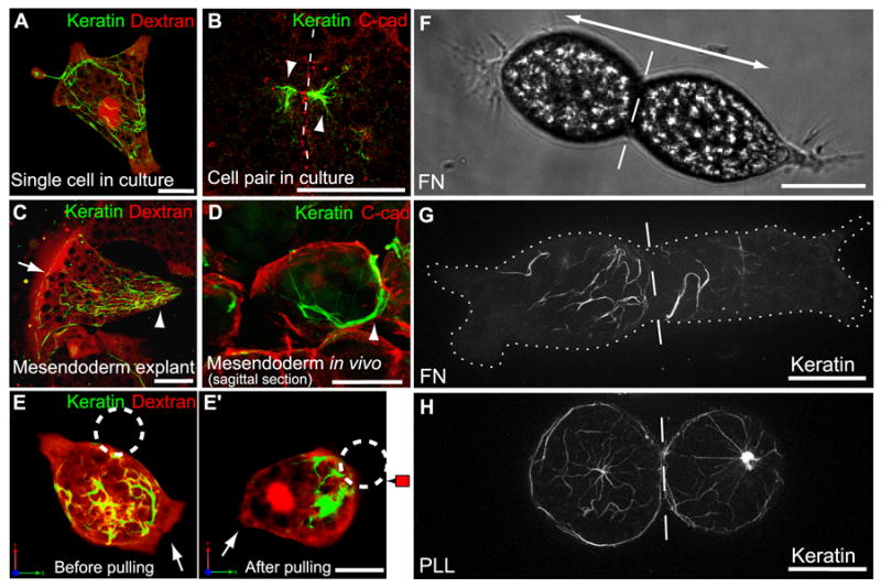
(A) Single cell on FN, labeled with Alexa555-dextran (red) and expressing GFP-XCK1(8) to visualize KIFs (green). (B) Pair of fixed mesendoderm cells immunostained for C-cadherin (red) and XCK1(8) (green). Dashed line, cell-cell boundary. (C) Cell within mesendoderm tissue explant on FN labeled with Alexa555-dextran (red) and expressing GFP-XCK1(8) (green). (D) Sagittal perspective of mesendoderm cell in bisected embryo immunostained for C-cadherin (red) and XCK1(8) (green). KIFs in posterior of polarized cells (arrowheads B-D) and along tissue leading edge (arrow in C). (E,E’) Single mesendoderm cell on FN labeled with Alexa555-dextran (red), expressing GFP-XCK1(8) (green). C-cadFc bead (dashed circle) attached to cell (E), then pulled for 20 min (E’). Arrows, leading edge protrusion. (F) Brightfield image of cell pair on FN, polarized in opposing directions (double arrow). (G,H) Cell pairs expressing GFP-XCK1(8), plated on FN (G) or PLL (H). Dashed line, cell-cell boundary. Cell borders outlined by dotted line in (G). All scale bars, 25μm. See also Movie S4.