
Figure 6. Requirement for PG and Keratin in Normal Mesendoderm In Vivo.
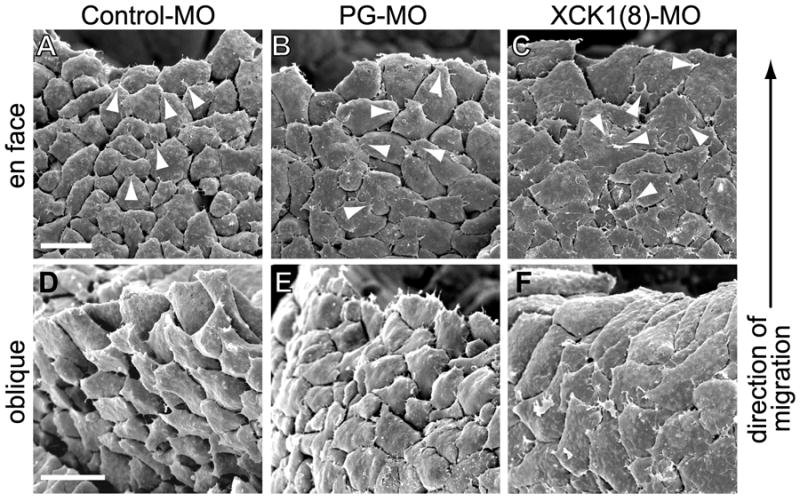
Scanning electron micrographs of Xenopus embryos from which the overlying blastocoel roof was removed to reveal the basal aspect of the underlying mesendoderm (as in Figure 1A). Leading edge mesendoderm cells and direction of migration in all images is toward top. Images were acquired of (A,D) control morpholino injected embryos, (B,E) PG morpholino injected embryos, and (C,F) XCK1(8) morpholino injected embryos. En face view of basal aspect shown in (A-C) and oblique view of the basal surface shown in (D-F). Arrowheads indicate a sampling of cell protrusions. Scale bars, 50μm.