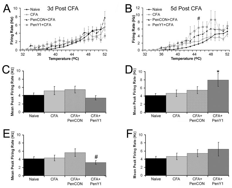
Figure 4. Firing rates of CPM neurons during heat stimuli before and after injection of CFA into the hairy hindpaw skin.
Firing rate per degree to heat stimuli in CPM neurons is unaffected by CFA at three (Panel A) and five (Panel B) days under all three conditions compared to naïves with the exception of the 45°C temperature at five days where inhibition of P2Y1 during inflammation was significantly higher than naives. However, this increase was not statistically different than CFA alone or PenCON plus CFA groups. #p value < 0.05 relative to naïves. When calculating maximal firing rate over the entire heat ramp, there was no difference in the maximal firing rate to heat stimuli at three days after CFA injection (Panel C) under any condition; however, inhibition of P2Y1 did cause a significant increase in the maximal firing rate of CPM neurons to heat stimuli (Panel D). * p value < 0.05 compared to naives. Removal of the TRPV1 immunopositive/peptidergic CPM neurons from these data reveal that increases in firing rate by inhibition of P2Y1 five days after CFA injection (Panel F) is due to this small population of CPM neurons. In fact at the three day time point, removal of these few CPM neurons reduced the maximal firing rate below that of the PenCON plus CFA group but this was not different than naives or CFA alone (Panel E). # p value < 0.05 relative to PenCON plus CFA.

