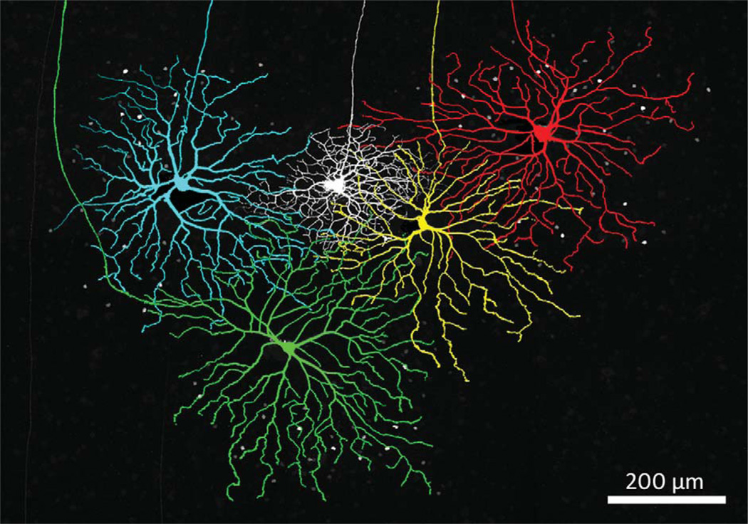
Figure 9.
Four Neurobiotin-injected coupled ON DS ganglion cells surrounding an ON-OFF DS ganglion cell (white) show the relative size of these two cell types. The ganglion cells were colored in Adobe Photoshop for clarity. The micrograph is constructed from 35 × 1.0 µm optical sections. These four ON DS ganglion cells are not necessarily nearest neighbors.

