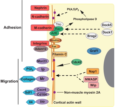
Fig. 6.
Genes and pathways associated with myoblast fusion in mice. The indicated proteins and pathways correspond to those, as discussed in the text, for which a role in fusion has been shown experimentally. Generally, these comprise cell-adhesion molecules and their associated adaptors, secreted molecules and their receptors that control cell migration, and molecules that signal to the actin cytoskeleton. For the most part, roles for these proteins have been identified through loss-of-function studies. Relationships indicated by broken lines are based on studies in other tissues, but have not yet been established in this system. Arf6, ADP ribosylation factor 6; Brag2, brefeldin A-resistant Arf GEF; CD164, cluster of differentiation 164; Cdc42, cell division cycle 42; Cxcr4, chemokine (C-X-C motif) receptor 4; Dock1 and Dock 5, dedicator of cytokinesis 1 and 5; Fak, focal adhesion kinase; Graf1, GTPase regulator associated with focal adhesion kinase; Il4, interleukin 4; Il4r, interleukin 4 receptor; Ip, prostacyclin receptor; Mr, mannose receptor; Mor23, mouse odorant receptor 23; Nap1, Nck-associated protein; NWASP, neural Wiskott-Aldrich syndrome protein; PGI2, prostacyclin; PI(4,5)P2, phosphatidylinositol 4,5-bisphosphate; Rac1, Ras-related C3 botulinum toxin substrate 1; SDF1, stromal-derived factor 1; Wip, WASp-interacting protein.

