Fig. 5.
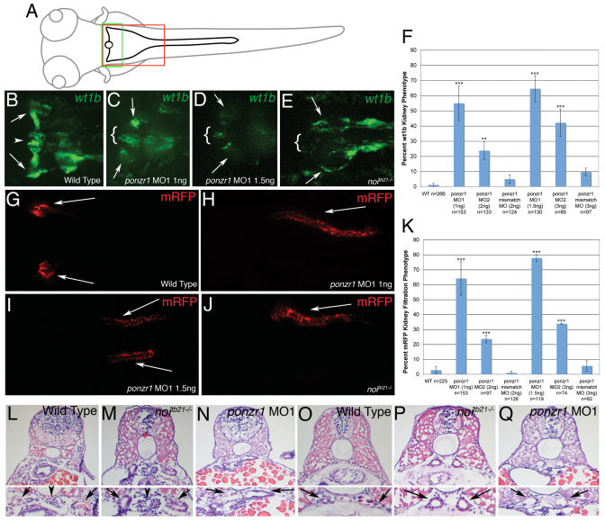
ponzr1 is required for anterior kidney form and function. (A) A drawing of a larval zebrafish at 3-4 dpf. The green box is the area of the larva seen in images B-E, and the red box is the area seen in images G-J. (B) Tg(wt1b:EGFP) larvae (Perner et al., 2007) mark podocytes surrounding the glomerulus (arrowhead) and the pronephric tubules (arrows). (C,D) ponzr1 morphants show a loss of fluorescence in the area surrounding the glomerulus, except at the midline (brackets). The pronephric tubular staining is lost (arrows) in a dose-dependent manner (D: 1 ng MO1; E: 1.5 ng MO1). (E) noitb21–/– mutant embryos exhibit a loss of fluorescence around the glomerulus (brackets) and the pronephric tubule fluorescence runs posteriorly instead of laterally (arrows). (F) Two ponzr1 MOs induce a similar phenotype that is ameliorated in mismatch control MO injections. (G) mRFP appears bilaterally in the curvature of the pronephric tubules (arrows) in control embryos due to glomerular filtration. (H-J) In ponzr1 morphants, mRFP is collected in a more posterior part of the tubules – sometimes only one pronephric tubule collects fluorescence and sometimes fluorescence is seen in both (H: 1 ng MO1; I: 1.5 ng MO1). A similar phenotype is seen in noitb21–/– mutant embryos (J). (K) Percentage of embryos with the filtration phenotype. (L-Q) Larvae at 4dpf that have been sectioned and H&E stained. (L) Uninjected embryos show a glomerulus (arrowhead) with two pronephric tubules (arrows). (M) noitb21–/– mutant embryo has a glomerulus (arrowhead) but shows dilated tubules (arrow). (N) ponzr1 morphants show dilated pronephric tubules (arrows) with no glomerulus. (O) The posterior pronephric tubules in wild-type embryos (arrows). noitb21–/– mutant (P) and ponzr1 morphant embryos (Q) both show dilated tubules (arrows). **P<0.01, ***P<0.001

